Inferior Meatus
Contents of Drilled Internal Auditory Canal | Neuroanatomy | The. 9 Pics about Contents of Drilled Internal Auditory Canal | Neuroanatomy | The : Posterior View of Medulla, Fourth Ventricle, Internal Auditory Meatus, Nasal cavity: Anatomy, structure, parts, blood supply | Kenhub and also Contents of Drilled Internal Auditory Canal | Neuroanatomy | The.
Contents Of Drilled Internal Auditory Canal | Neuroanatomy | The
 www.neurosurgicalatlas.com
auditory neurosurgicalatlas correlation
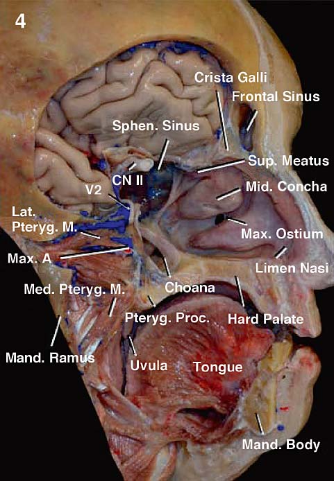
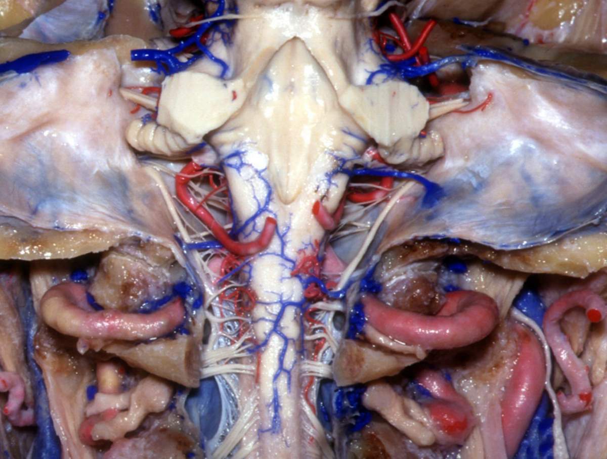
Anatomy Of The Nasal Cavity And Paranasal Sinuses | Neupsy Key
 neupsykey.com
cavity nasal anatomy sinuses maxillary paranasal
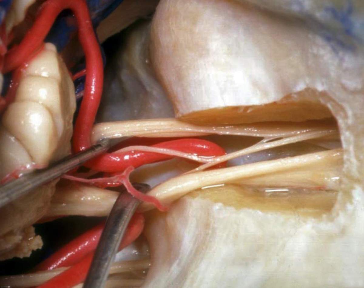
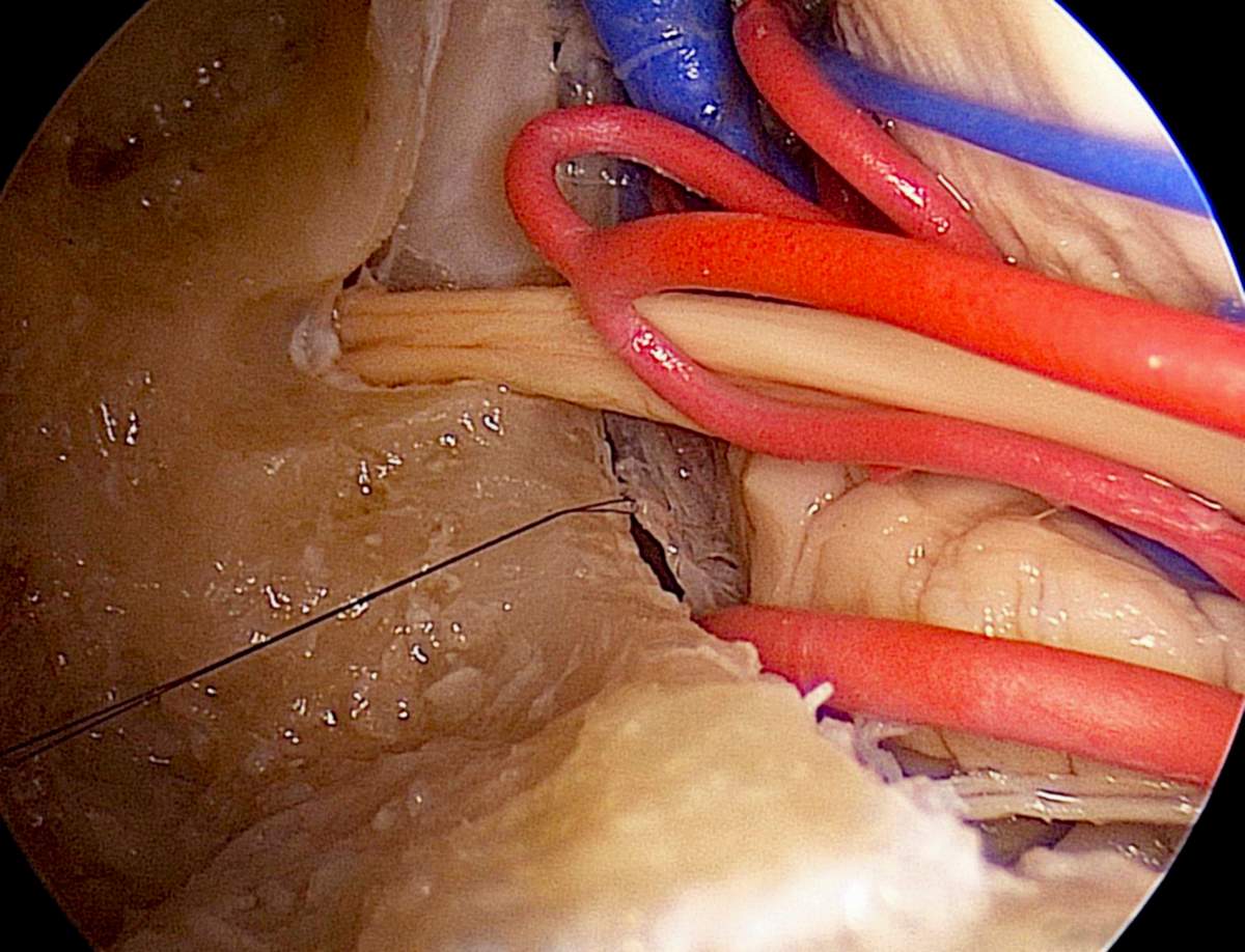
Posterosuperior View Of Left Cerebellopontine Angle | Neuroanatomy
 www.neurosurgicalatlas.com
posterosuperior cerebellopontine neurosurgicalatlas correlation
Posterior View Of Medulla, Fourth Ventricle, Internal Auditory Meatus
 www.neurosurgicalatlas.com
meatus auditory internal posterior medulla foramen ventricle fourth jugular neurosurgicalatlas
The Endoscopic Anatomy Of The Middle Ear Approach To The Fundus Of The
 thejns.org
ear anatomy middle endoscopic internal process fig fundus canal acoustic approach volume issue
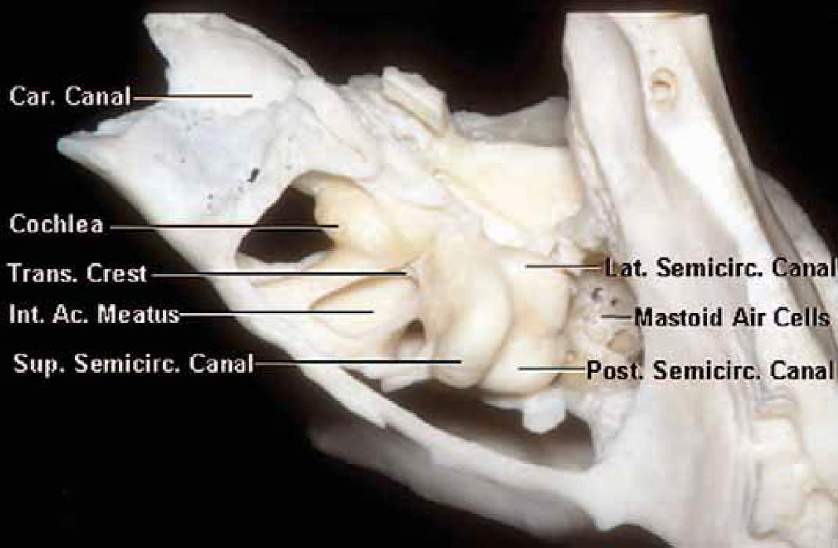
The Osseous Capsule Of The Cochlea, Semicircular Canals, And Internal
 www.neurosurgicalatlas.com
semicircular canals cochlea meatus osseous neurosurgicalatlas
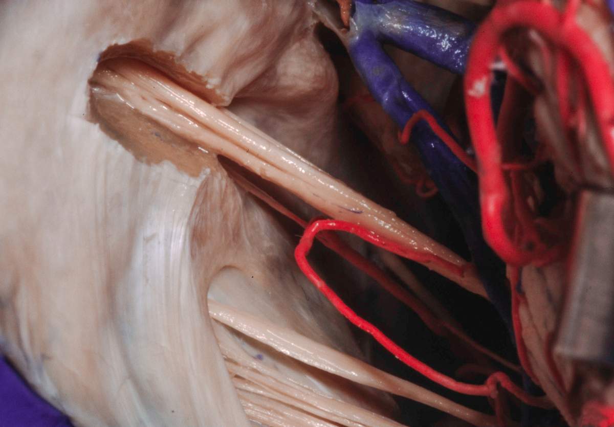
Magnified Endoscopic View Of Porus Of Right Internal Auditory Meatus
 www.neurosurgicalatlas.com
meatus internal auditory right endoscopic magnified porus neurosurgicalatlas
Posterior View Of The Medulla And Spinal Cord | Neuroanatomy | The
 www.neurosurgicalatlas.com
spinal cord medulla posterior neuroanatomy neurosurgicalatlas
Nasal Cavity: Anatomy, Structure, Parts, Blood Supply | Kenhub
nasal anatomy superior meatus cavity nasi kenhub inferior structure conchae supply into middle called blood bony parts
Ear anatomy middle endoscopic internal process fig fundus canal acoustic approach volume issue. Spinal cord medulla posterior neuroanatomy neurosurgicalatlas. The endoscopic anatomy of the middle ear approach to the fundus of the