foot x ray anatomy
Radiographic Anatomy of the Skeleton: Elbow -- Lateral View, Labelled. 9 Pictures about Radiographic Anatomy of the Skeleton: Elbow -- Lateral View, Labelled : Osseous injuries of the foot: an imaging review. Part 1: the forefoot, Radiographic Anatomy of the Skeleton: Ankle -- Anteroposterior (AP and also Ollier's disease | Image | Radiopaedia.org.
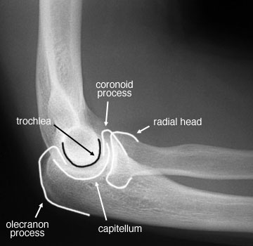
Radiographic Anatomy Of The Skeleton: Elbow -- Lateral View, Labelled
 uwmsk.org
elbow lateral joint ray anatomy labelled skeleton radiology radiographic process coronoid olecranon trochlea ap unlabelled ulna radial student head humerus
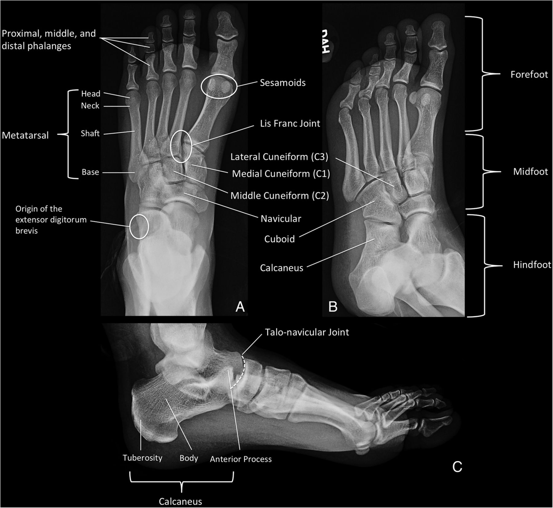
Osseous Injuries Of The Foot: An Imaging Review. Part 1: The Forefoot
 emj.bmj.com
forefoot foot oblique ap views figure injuries osseous distal emermed emj bmj powerpoint imaging tab open
Ollier's Disease | Image | Radiopaedia.org
 radiopaedia.org
ollier disease orthobullets foot radiology radiopaedia pathology
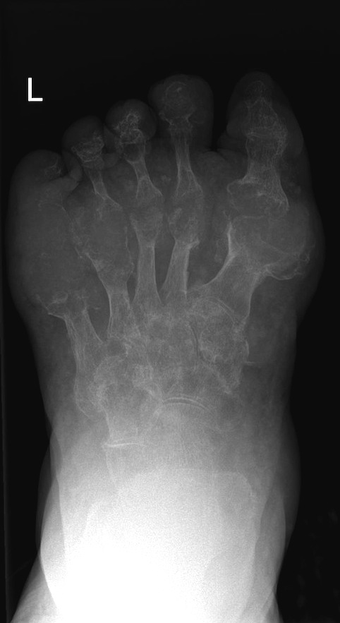
Gout - Radiology At St. Vincent's University Hospital
 www.svuhradiology.ie
gout radiology
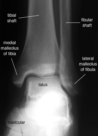
Radiographic Anatomy Of The Skeleton: Ankle -- Anteroposterior (AP
 uwmsk.org
ankle labelled ap anatomy skeleton radiology anteroposterior radiographic mortise foot student joint knee elbow
Xray Projections Of The Calcaneus - YouTube
 www.youtube.com
ray calcaneus xray positioning
Gout | Image | Radiopaedia.org
 radiopaedia.org
gout foot radiopaedia oblique case version intraosseous geodes lesion
Radiographic Anatomy - Hand Lateral | Radiographic Anatomy | Radiology
 www.pinterest.com
hand anatomy lateral ray radiographic xray radiology medical radiograph wikiradiography bones wrist carpal bone labels lumbar radiography technology rad tech
Congenital Talipes Equinovarus | Image | Radiopaedia.org
 radiopaedia.org
talipes equinovarus congenital foot club bilateral radiopaedia right feet case version radiology
Radiographic anatomy of the skeleton: ankle -- anteroposterior (ap. Gout foot radiopaedia oblique case version intraosseous geodes lesion. Hand anatomy lateral ray radiographic xray radiology medical radiograph wikiradiography bones wrist carpal bone labels lumbar radiography technology rad tech