foot joints anatomy
Foot X-rays taken at acute presentation (AP and lateral views. 9 Images about Foot X-rays taken at acute presentation (AP and lateral views : {Bones of the foot separated | John The Bodyman, Osseous injuries of the foot: an imaging review. Part 1: the forefoot and also Foot X-rays taken at acute presentation (AP and lateral views.
Foot X-rays Taken At Acute Presentation (AP And Lateral Views
 www.researchgate.net
demonstrating
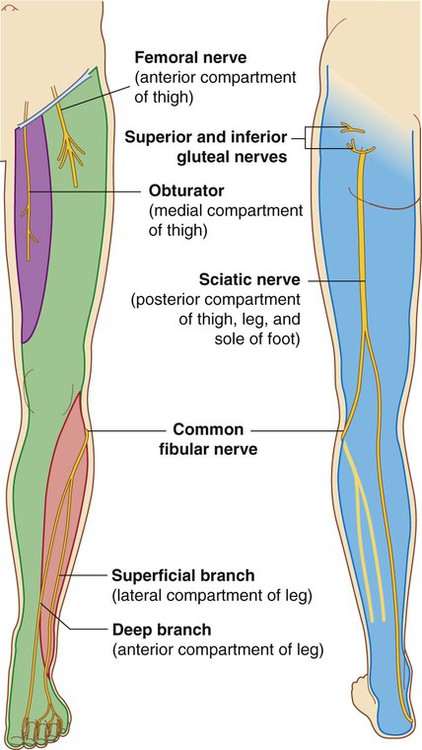
Lower Limb: Bones, Muscles, Joints & Nerves » How To Relief
 www.howtorelief.com
lower limb nerves muscles nerve anatomy innervation joints motor muscle body major extremity sciatic anterior inferior bones cutaneous posterior regions
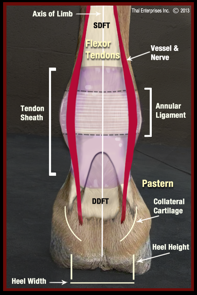
Vitals & Anatomy - Horse Side Vet Guide
 horsesidevetguide.com
horse limb equine anatomy joint rear lower fetlock horses swollen ankle lameness tendon sheath swelling side horsesidevetguide ligament injury vet
Joints Of The Upper Limb ~ Anatomy For MSP
 aftabphysio.blogspot.com
wrist joint joints hand arm anatomy ellipsoidal body complex daviddarling info between msp services
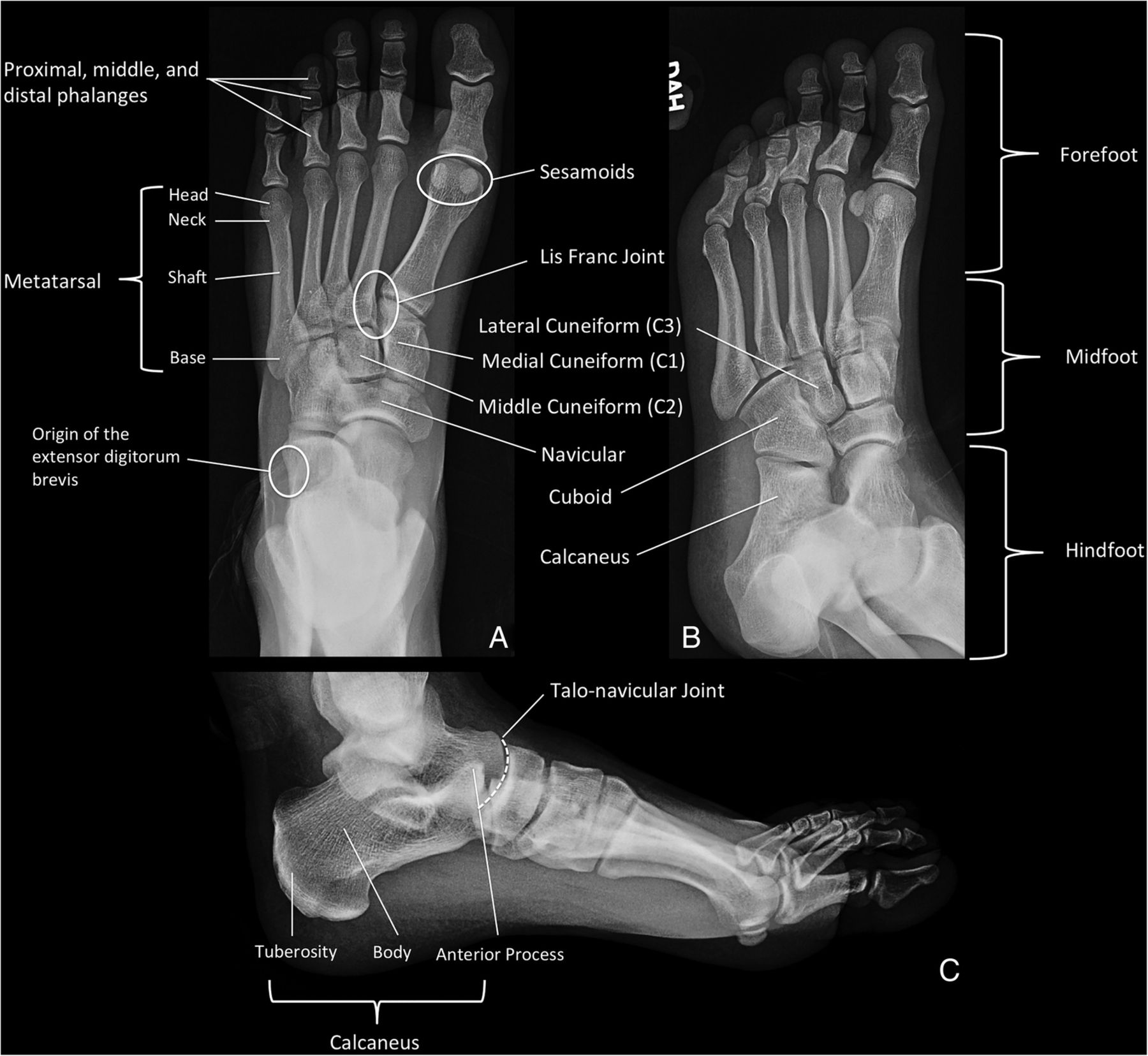
Osseous Injuries Of The Foot: An Imaging Review. Part 1: The Forefoot
 emj.bmj.com
forefoot foot oblique ap views figure injuries osseous distal emermed emj bmj powerpoint imaging tab open
Surface Anatomy
hindfoot position knee
{Bones Of The Foot Separated | John The Bodyman
 johnthebodyman.com
bones foot labeled bone label ankle separated labled human johnthebodyman
Foot Anatomy And Biomechanics - Foot & Ankle - Orthobullets
 www.orthobullets.com
eversion inversion orthobullets foot ankle anatomy joint tarsal transverse motion dorsiflexion biomechanics talonavicular plantar topic upload
The Anatomy Of A Foot
 www.orangeinsoles.com
forefoot
Bones foot labeled bone label ankle separated labled human johnthebodyman. Lower limb nerves muscles nerve anatomy innervation joints motor muscle body major extremity sciatic anterior inferior bones cutaneous posterior regions. Foot anatomy and biomechanics