fetal hip bone anatomy
Ultrasound Evaluation of Normal Fetal Anatomy | Radiology Key. 9 Pictures about Ultrasound Evaluation of Normal Fetal Anatomy | Radiology Key : Hip joint: Bones, movements, muscles | Kenhub, Principles of Bone Grafting for Osteonecrosis of the Hip | Radiology Key and also Ultrasound Evaluation of Normal Fetal Anatomy | Radiology Key.
Ultrasound Evaluation Of Normal Fetal Anatomy | Radiology Key
 radiologykey.com
fetal anatomy
Untitled Document [bio.sunyorange.edu]
![Untitled Document [bio.sunyorange.edu]](http://bio.sunyorange.edu/updated2/comparative_anatomy/anat_3/hip_files/49c.jpg) bio.sunyorange.edu
bio.sunyorange.edu
hip chimp human pelvis anatomy chimpanzee updated2 hips evolution thinking between 49c homo leg sunyorange bio edu
Compact Bone Structure Microscopic Level – Medical Stock Images Company
 www.medicalstockimages.net
bone compact structure
Hip Joint: Bones, Movements, Muscles | Kenhub
hip joint kenhub anatomy bone human ligaments femur muscles bones movements ventrolateral
MediVisuals Normal GI Tract Development Medical Illustration
 medivisuals1.com
gi tract development normal medivisuals1
Principles Of Bone Grafting For Osteonecrosis Of The Hip | Radiology Key
 radiologykey.com
bone hip grafting osteonecrosis principles
Childbirth Demonstration Pelvis Skeleton Model With Fetal Skull - 3B
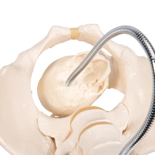 www.3bscientific.co.uk
childbirth fetal pelvis parto bacino illustrare becken l30 demonstratie bevalling 3bscientific
Femur Bone Anatomy: Proximal, Distal And Shaft | Kenhub
tuberculum femur kenhub adductor tubercle pelvis femoris proximal distal
Sacrum: Anatomy And Clinical Aspects | Kenhub
sacrum posterior kenhub anatomy bony landmarks
Hip joint: bones, movements, muscles. Ultrasound evaluation of normal fetal anatomy. Medivisuals normal gi tract development medical illustration